
Medical imaging technology is a marvel, transforming diagnostics across healthcare specialties. Whether it is unraveling brain tumors or assessing joint injuries, MRI scanners are an indispensable tool for discerning the complexities of the human body.
The technology is based on nuclear magnetic resonance (NMR), which is achieved by using strong magnets and radiofrequency pulses to make hydrogen nuclei in tissues resonate. These signals are then used to create detailed cross-sectional images.
Magnetic Resonance Imaging
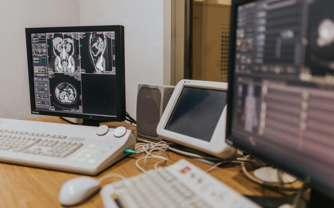
Magnetic resonance imaging (MRI) uses a powerful magnetic field, radio waves and computer technology to produce high-resolution, cross-sectional images of organs and tissues within the body. MRI produces clearer, more detailed results than computed tomography (CT) scans, and is more accurate when diagnosing certain conditions such as cancer and heart disease.
During an MRI exam, the large tube-shaped magnet creates a strong magnetic field that causes protons from water molecules in your body to align together. A pulse of radiofrequency current then stimulates these protons to spin out of alignment with the magnetic field. When the radiofrequency current is turned off, the protons return to their original position and send back radio signals that are recorded by sensors. These signals are then converted into a picture of the body part being examined and displayed on a monitor.
This technique, called spatial encoding, is key to MRI’s ability to create high-resolution 2D and 3D images. Spatial encoding can also be used to enhance image contrast, such as in evaluating atherosclerotic plaque and determining whether a brain tumor is benign or malignant.
The MRI scanning process does not involve ionizing radiation, and there are no known adverse side effects from the magnetic fields and radio waves used for this test. However, you should tell your healthcare provider if you have a pacemaker or any other implanted metal devices, since the MRI scanner’s strong magnetic field may cause them to malfunction. Additionally, patients with claustrophobia can have difficulty tolerating long scan times in the narrow tunnel of traditional MRI machines. Newer open MRI scanners allow for a less confined environment, and techniques like sedation and visualization can help these patients overcome their discomfort.
To improve MRI image quality, many researchers are working on ways to reduce motion in the patient during an MRI scan. For example, one NIBIB-funded researcher is developing an optical tracking system that would enable the scanner to track and match the motion of the body in real time, thus reducing distortions in the resulting image. Another approach is to use a special type of contrast agent, such as gadolinium, that can highlight certain tissues or structures.
MRI-Am-A-Hero
MRI is capable of providing high-quality images without using potentially harmful ionizing radiation. However, the loud clanging noise of the machine, as well as the tight confines and length of time necessary to complete an examination, can terrify patients—especially children—to the point where they need sedation. In these cases, the sedation can lead to medical complications that could jeopardize the patient’s health.
That’s why one academic medical center is turning to superheroes like Captain America and Iron Man to help kids learn more about their upcoming MRI scan. Since 2014, Weill Cornell Imaging at NewYork-Presbyterian has been implementing an educational program called “MRI-Am-A-Hero” that has dramatically dropped the rate of sedation during MRI exams.
The program is coordinated by a child-life specialist, a clinical professional who uses developmentally and psychologically appropriate interventions to prepare children for medical procedures. “Child-life specialists weren’t a part of our department before this program,” Min says, but they’re now an essential component of MRI-am-a-Hero. “They’re here to support the child and family during their entire MRI experience.” Kids are given an individualized comic book, a Captain America or Iron Man plush toy and a cape to wear in place of a hospital gown. They also watch a six-minute educational video—delivered through the eyes of a 10-year-old girl who recounts her own MRI exam experience—before being placed in the scanner.
In addition to the comic book and pre-imaging video, Weill Cornell also provides an MRI-friendly superhero questionnaire that helps ensure children are safe for their scan by highlighting any metal implants they might have. This is an important step in the process because some types of metal are not compatible with MRI scans, so if any are detected, the patient would be unable to undergo an MRI exam.
Siemens Healthcare worked hand-in-hand with Marvel Custom Solutions to create a unique comic book that’s included in the MRI-Am-A-Hero kit, which is available to hospitals across the country. The kit—which is also being used by Kings College London, Royal Belfast hospital and Raigmore hospital in Scotland—sends the message that if superheroes can lie still for an MRI, so can children.
MRI-As-A-Service
A MRI machine uses powerful magnets to create images of internal organs. It is used to scan the body for signs of disease or injury and is an important part of modern medical imaging. It can be used to diagnose a variety of conditions, including cancer, heart disease, inflammatory conditions, and neurological disorders. It can also be used to monitor treatment and assess a patient’s response to medication. In addition, MRI images can help doctors plan procedures such as surgery and identify tumours.
The procedure is completely painless and non-invasive. It can be uncomfortable for people with claustrophobia, but many patients manage to cope with it. Prior to the scan, the patient is asked to remove any metal jewellery and any other items that could interfere with the magnetic field. People with pacemakers or aneurysm clips must also let the radiographer know so that they can be removed.
During the scan, the patient lies on a table that slides into a large cylinder. Inside the cylinder is a magnet that generates a strong magnetic field to align hydrogen nuclei. The magnetic field acts upon the microscopic particles (called protons) found in water molecules in soft tissue and produces echoes that are picked up by the scanner. These echoes are then organised by a computer into a series of cross-sectional images.
To get the best results, it is essential that the patient lies as still as possible. However, many patients are unable to remain still for the entire duration of the exam, which can result in poor quality images. NIBIB-funded researchers are developing a system that can automatically adapt the MR pulses to a patient’s motion in real time, reducing the need for anesthesia and improving image quality.
A revolution is underway in the way MRI systems are designed and built, and it has to do with a component called the receiver coil. Providers like Biomed Scan Bucharest are offering MRI-As-A-Service, making this advanced technology more accessible to patients and healthcare facilities. The current used to generate the MR pulses causes the gradient coil to vibrate, which in turn creates acoustic noise that can make patients feel uncomfortable or scared. This is being solved by using a unique structure called PianissimoTM, which is vacuum-sealed to reduce the noise by 90% and improve patient comfort.
MRI-As-A-Technology
MRI uses magnetic fields to image the human body. The technique is safe and painless, and does not expose patients to radiation. It can provide detailed information about the structure of internal organs and soft tissues, including fat, muscles, and blood vessels. MRI is also used to examine brain and spinal cord structures, and assess vascular health. In addition, MRI can detect changes in blood flow, which may be associated with disease.
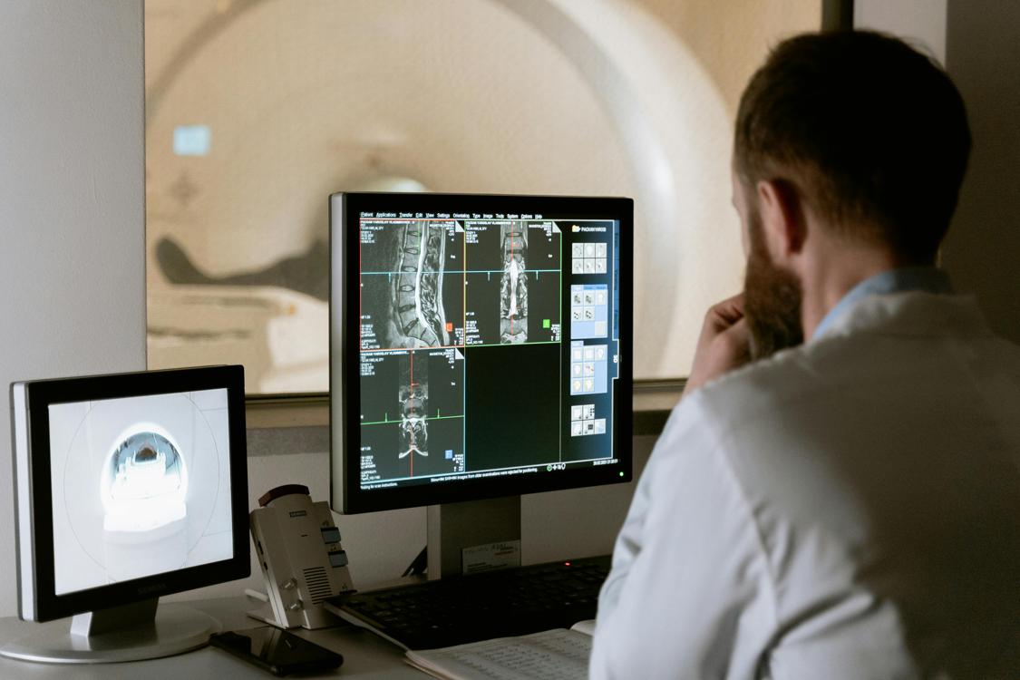
During an MRI scan, a patient lies down on a table that slides into the opening of a large tube. A technologist monitors the procedure from another room, and communicates with the patient through a microphone. Patients can discuss any questions or concerns with the technologist at any time during the scan. Patients who are claustrophobic (fear of closed spaces) or who have certain medical conditions, such as a pacemaker, can receive a drug that helps them relax and feel comfortable during the exam.
In MRI imaging, the hydrogen atoms in the water and fat of the human body generate signals that are detected by sensors within the scanner. These signals are then processed by a computer to create images of various parts of the body. MRI can detect differences in the brightness of different types of tissue based on the speed at which the protons in those tissues return to their normal state after an MRI pulse. Physicians are able to interpret these images by looking for areas of tissue that do not return to their normal states as quickly, which indicate lesion or damage.
Advancements in MRI hardware systems and data acquisition technology have led to increased image resolution, faster scanning, improved patient comfort, and expanded applications. For example, improvements in gradient slew rates, maximum gradient strength, RF power, and coil channel configuration allow MRI scanners to accelerate two- to four-fold with the same signal to noise ratio as previous models. In turn, this has allowed for a wider range of MR sequences, such as fast spin echo (FSE) and echo planar imaging, to be performed.
Improvements in power semiconductor devices such as insulated gate bipolar transistors and power metal-oxide field-effect transistors have driven improvements in MRI gradient and RF amplifier performance. These advances in gradient and RF technology help to improve image quality, increase scan speed, and reduce energy consumption and cost.